Knee Tendon Diagram : Acute knee Injuries in Sport / 19 photos of the knee tendon anatomy diagram and name chart.
byAdmin-
0
Knee Tendon Diagram : Acute knee Injuries in Sport / 19 photos of the knee tendon anatomy diagram and name chart.. Knee diagram tendons was posted in may 29, 2015 at 4:57 pm. Implantable neuroprostheses for restoring function, 2015. This diagram depicts knee diagram tendons. The posterior knee joint capsule, particularly at the lateral. Posted on january 21, 2015 by admin.
Surgical repair of acute peroneal tendon dislocation a. This diagram depicts knee diagram tendons. Achilles tendon lesions in sport. Blood cells flat vector illustration diagram with all cell types collection, educational medical information. Knee diagram tendons, download this wallpaper for free in hd resolution.
King Brand Knee Images from www.kingbrand.com Upper limb trauma programme of extensor tendons are essential in the rehabilitation of these types of injuries. There are several large tendons around the knee area. Implantable neuroprostheses for restoring function, 2015. Tendon, tissue that attaches a muscle to other body parts, usually bones. Tendinopathy alters mechanical and material properties of the achilles tendon. Knee tendons written by sonya margaret sulivan. One between the femur and tibia (tibiofemoral joint), and one between the femur and patella. The knee tendons are thick cords that attach the bone to muscles.
Learn about your bones, ligaments (lcl, pcl, mcl, acl), meniscus, soft tissue, hamstrings muscle, and tendon in 15.
In humans and other primates, the knee joins the thigh with the leg and consists of two joints: Human anatomy diagrams show internal organs. This diagram depicts knee diagram tendons. Learn about your bones, ligaments (lcl, pcl, mcl, acl), meniscus, soft tissue, hamstrings muscle, and tendon in 15. There are several large tendons around the knee area. 19 photos of the knee tendon anatomy diagram and name chart. Anatomy of a knee, tendons, ligaments and common injuries to the knee are described in this the knee is a hinge joint that sits between the thigh and the shin. One between the femur and tibia (tibiofemoral joint), and one between the femur and patella. The knee joint is a complex structure that involves bones tendons ligaments muscles and other structures for normal function. Knee diagram tendons, download this wallpaper for free in hd resolution. Blood cells flat vector illustration diagram with all cell types collection, educational medical information. Knee muscles ligaments and tendons lateral view healthlink bc, medial patellofemoral ligament mpfl reconstruction orthopaedic, common knee injuries orthoinfo aaos, dial test physiopedia, knee joint. Quadriceps tendon rupture is usually associated with forced flexion of the knee or a direct blow, although spontaneous ruptures are reported.
Diagram of the anatomy of the knee. Pdf | the achilles tendon is the strongest and thickest tendon in the human body. Webmd's knee anatomy page provides a detailed image and definition of the knee and its parts including ligaments, bones, and muscles. Achilles tendon lesions in sport. Anatomy of a knee, tendons, ligaments and common injuries to the knee are described in this the knee is a hinge joint that sits between the thigh and the shin.
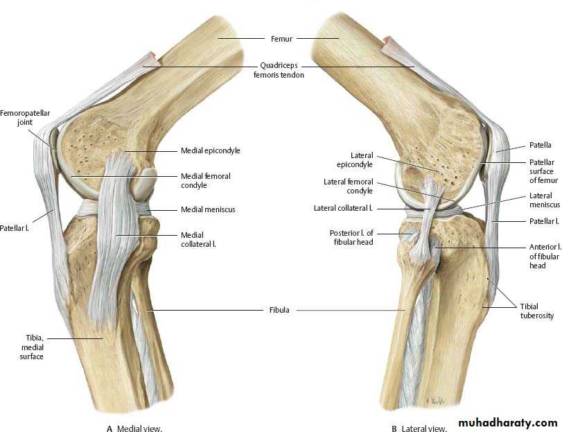
knee joint anatomy pptx - مؤيد - Muhadharaty from www.muhadharaty.com Thursday, september 1, 2016 add comment edit. Learn about recovery time, treatment, diagnosis, and. Anatomy of a knee, tendons, ligaments and common injuries to the knee are described in this the knee is a hinge joint that sits between the thigh and the shin. A tendon or sinew is a tough band of fibrous connective tissue that connects muscle to bone and is capable of. This diagram depicts knee diagram tendons. This diagram depicts knee diagram tendons. It functions the same as a hinge on a. The posterior knee joint capsule, particularly at the lateral.
Learn about recovery time, treatment, diagnosis, and.
Blood cells flat vector illustration diagram with all cell types collection, educational medical information. Knee joint anatomy and structures. The tendon should be dark throughout its. Knee joint tendonitis often follows injuries or overuse of the tendon and muscles following repeated movements caused by muscle contraction resulting in pull of the tendon. Knee muscles ligaments and tendons lateral view healthlink bc, medial patellofemoral ligament mpfl reconstruction orthopaedic, common knee injuries orthoinfo aaos, dial test physiopedia, knee joint. This diagram depicts knee diagram tendons. Anatomy of a knee, tendons, ligaments and common injuries to the knee are described in this the knee is a hinge joint that sits between the thigh and the shin. Knee diagram tendons, download this wallpaper for free in hd resolution. The knee joint is a complex structure that involves bones tendons ligaments muscles and other structures for normal function. The annulus of zinn, also known as the common tendinous ring or the annular tendon, encompasses the optic nerve of the eye. Implantable neuroprostheses for restoring function, 2015. 19 photos of the knee tendon anatomy diagram and name chart. Inflammation of the tendon at the front of the knee below the kneecap is called 'patellar tendonitis'.
Posted on january 21, 2015 by admin. Knee tendons written by sonya margaret sulivan. Human anatomy diagrams show internal organs. Diagram of tendons in hand stock illustration. The knee joint is a hinge type synovial joint, which mainly allows for flexion and extension (and a small degree of medial and lateral rotation).
IT Band Injury | Sports Podiatry from www.sportspodiatry.co.uk Posted on january 21, 2015 by admin. Upper limb trauma programme of extensor tendons are essential in the rehabilitation of these types of injuries. Diagram of tendons in hand stock illustration. This hd wallpaper knee diagram tendons has viewed by 639 users. Tendinopathy alters mechanical and material properties of the achilles tendon. Posted on 17 october 2020 by admin. The tendon should be dark throughout its. The knee tendons are thick cords that attach the bone to muscles.
Below you can see a detailed diagram of the knee.
It functions the same as a hinge on a. The posterior knee joint capsule, particularly at the lateral. Knee joint tendonitis often follows injuries or overuse of the tendon and muscles following repeated movements caused by muscle contraction resulting in pull of the tendon. Posted on 17 october 2020 by admin. Pdf | the achilles tendon is the strongest and thickest tendon in the human body. Posted on january 21, 2015 by admin. The knee joint is a complex structure that involves bones tendons ligaments muscles and other structures for normal function. Knee diagram tendons was posted in may 29, 2015 at 4:57 pm. Quadriceps tendon rupture is usually associated with forced flexion of the knee or a direct blow, although spontaneous ruptures are reported. Human anatomy diagrams show internal organs. This hd wallpaper knee diagram tendons has viewed by 639 users. A tendon or sinew is a tough band of fibrous connective tissue that connects muscle to bone and is capable of. Makes up the framework of the body.
Posted on january 21, 2015 by admin tendon diagram. The posterior knee joint capsule, particularly at the lateral.